Deviated Septum

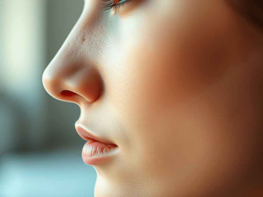
The bone and cartilage that divides the inside of the nose in half is called the nasal septum. The bone and cartilage are covered by a special skin called a mucous membrane that has many blood vessels in it.

Ideally, the left and right nasal passageways are equal in size. However, it is estimated that as many as 80 percent of people have a nasal septum that is off-center. This is called a deviated septum, which may or may not cause certain symptoms.

What Are the Symptoms of a Deviated Septum?
The most common symptom from a badly deviated or crooked septum is difficulty breathing through the nose, which is usually worse on one side. In some cases, a crooked septum can interfere with sinus drainage and cause repeated sinus infections. You may experience one or more of the following:

Difficulty breathing through one or both nostrils
Nosebleeds
Sinus infections
Noisy breathing during sleep in infants and young children
Mouth-breathing during sleep in adults

What Causes a Deviated Septum?

Injury or trauma to the nose can cause the septum to become deviated or crooked. However, even people with normal growth and development, and without a history of injury, trauma, or broken nose, can have a deviated septum.

What Are the Treatment Options?

Discuss your symptoms and any known nose damage or surgeries with your primary care provider or an ENT (ear, nose, and throat) specialist, or otolaryngologist. They will examine your nose inside and out, and might recommend additional tests based on your individual needs. When there is clearly a crooked/deviated septum, and the symptoms are severe enough to warrant intervention, the ENT specialist may suggest surgery as an option if medical treatment fails.

Septoplasty is the preferred surgical treatment to correct a deviated septum. This procedure is typically not performed on young children, unless the problem is severe, because facial growth and development are still occurring. Septoplasty is a surgical procedure that is usually performed through the nostrils, so there is no bruising or outward sign of surgery; however, each case is different and special techniques may be required depending on the individual patient.

The time required for the septoplasty operation averages about one- to one-and-a-half hours, depending on the type of deformity. It can be done with a local or a general anesthetic, usually on an outpatient basis. During the surgery, badly deviated portions of the septum may be removed entirely, or they may be readjusted and reinserted into the nose. Surgery may be combined with a rhinoplasty that changes the outward shape of the nose; in this case swelling and bruising may occur. Septoplasty may also be combined with sinus surgery.

Are There Related Factors or Conditions?

  • Inferior turbinate hypertrophy—turbinates are finger-like structures in your nose that warm and moisten the air you breathe, and sometimes the lower ones can get too big
  • Concha bullosa of the middle turbinate—this is when one of the turbinates next to your sinus openings gets a big air bubble in it
  • Nasal valve collapse (internal or external)
  • Sinusitis (acute, recurrent, chronic)
  • Headaches (contact point)
  • External nasal deformity (change in the shape of the nose)
  • Decreased sense of smell

Are There Potential Dangers or Complications?

Sometimes a deviated septum may lead to repeated nosebleeds. If the blockage is severe, it may force mouth-breathing at night, which can worsen sleep disorders. However, potential complications from septoplasty (surgery) can include:

  • Anesthesia complications
  • Bleeding
  • Infection
  • Creation of a hole connecting the right and left sides of the nasal cavity (called a septal perforation)
  • Numbness of the upper teeth and nose
  • Cerebrospinal fluid leak (extremely rare)
  • Change in the external shape of the nose

What Questions Should I Ask My Doctor?

  1. I’ve been experiencing some of these symptoms. Is it possible that I have a deviated septum and, if so, how severe do you think it is?
  2. Is there anything about my nose that might be interfering with my breathing?
  3. Are there any other conditions that may be contributing to my nasal congestion/obstruction?

Pediatric Sleep-disordered Breathing

Pediatric sleep-disordered breathing (SDB) is a general term for breathing difficulties during sleep. SDB can range from frequent loud snoring to obstructive sleep apnea (OSA), a condition where part, or all, of the airway is blocked repeatedly during sleep.

When a child’s breathing is disrupted during sleep, the body thinks the child is choking. The heart rate increases, blood pressure rises, the brain is aroused, and sleep is disrupted. Oxygen levels in the blood can also drop.

Approximately 10 per cent of children snore regularly, and about two to four per cent of children experience OSA. Recent studies indicate that mild SDB or snoring may cause many of the same problems as OSA in children.

What Are the Symptoms of Pediatric SDB?
Potential symptoms and consequences of untreated pediatric SDB may include:

Snoring—The most obvious symptom of SDB is loud snoring that is present on most nights. The snoring can be interrupted by a complete blockage of breathing, with gasping and snorting noises associated with waking up from sleep. Loud snoring can also become a significant social problem if a child shares a room with siblings, or at sleepovers and summer camp.
Irritability—A child with SDB may become irritable, sleepy during the day, or have difficulty concentrating in school. He or she may also display busy or hyperactive behaviour.
Bedwetting—SDB can cause increased urine production at night, which may lead to bedwetting (also called enuresis).
Learning difficulties—Children with SDB may become moody and disruptive, or not pay attention, both at home and at school. SDB can also be a contributing factor to attention deficit disorders in some children.
Slow growth—Children with SDB may not produce enough growth hormone, resulting in abnormally slow growth and development.
Cardiovascular difficulties—OSA can be associated with an increased risk of high blood pressure, or other heart and lung problems.
Obesity—SDB may cause the body to have increased resistance to insulin, and daytime fatigue can lead to decreased physical activity. These factors can contribute to obesity.
What Causes Pediatric SDB?
A common physical cause of airway narrowing contributing to SDB is enlarged tonsils and adenoids. Overweight children are at increased risk for SDB because fat deposits around the neck and throat can also narrow the airway. Children with abnormalities involving the lower jaw or tongue, or neuromuscular deficits such as cerebral palsy, have a higher risk of developing SDB.

How is Sleep Apnea Diagnosed?

If you notice any of the symptoms described in this article, have your child checked by an ENT (ear, nose, and throat) specialist, or otolaryngologist. Sometimes physicians will make a diagnosis of SDB based on history and physical examination. In other cases, like children suspected of having severe OSA due to craniofacial syndromes, morbid obesity, neuromuscular disorders, or for children less than three years old, additional testing such as a sleep test may be recommended.

The sleep study, or polysomnography (PSG), is an objective test for SDB. Wires are attached to the head and body to monitor brain waves, muscle tension, eye movement, breathing, and the level of oxygen in the blood. The test is not painful and is generally performed in a sleep laboratory or hospital. Sleep tests occasionally produce inaccurate results, especially in children. Borderline or normal sleep test results may still result in a diagnosis of SDB based on parental observations and clinical evaluation.

What Causes Pediatric SDB?
A common physical cause of airway narrowing contributing to SDB is enlarged tonsils and adenoids. Overweight children are at increased risk for SDB because fat deposits around the neck and throat can also narrow the airway. Children with abnormalities involving the lower jaw or tongue, or neuromuscular deficits such as cerebral palsy, have a higher risk of developing SDB.

How is Sleep Apnea Diagnosed?

If you notice any of the symptoms described in this article, have your child checked by an ENT (ear, nose, and throat) specialist, or otolaryngologist. Sometimes physicians will make a diagnosis of SDB based on history and physical examination. In other cases, like children suspected of having severe OSA due to craniofacial syndromes, morbid obesity, or neuromuscular disorders, or for children less than three years old, additional testing such as a sleep test may be recommended.

The sleep study, or polysomnography (PSG), is an objective test for SDB. Wires are attached to the head and body to monitor brain waves, muscle tension, eye movement, breathing, and the level of oxygen in the blood. The test is not painful and is generally performed in a sleep laboratory or hospital. Sleep tests occasionally produce inaccurate results, especially in children. Borderline or normal sleep test results may still result in a diagnosis of SDB based on parental observations and clinical evaluation.

What Are the Treatment Options?
Enlarged tonsils and adenoids are a common cause of SDB. Surgical removal of the tonsils and adenoids, called tonsillectomy and adenoidectomy (T&A), is generally considered the first-line treatment for pediatric SDB if the symptoms are significant, and the tonsils and adenoids are enlarged. Of the more than 500,000 pediatric T&A procedures performed in the United States each year, the majority treat SDB. Many children with OSA show both short- and long-term improvement in their sleep and behavior after T&A.

Not every child with snoring needs to undergo T&A. If the SDB symptoms are mild or intermittent, academic performance and behaviour is not an issue, the tonsils are small, or the child is near puberty (because tonsils and adenoids often shrink at puberty), it may be recommended that a child with SDB be watched conservatively and treated surgically only if symptoms worsen.

Recent studies have shown that some children have persistent SDB after T&A. A post-operative sleep study may be necessary, especially in children with persistent symptoms or increased risk factors for persistent apnea after T&A such as obesity, craniofacial anomalies or neuromuscular problems. Additional treatments such as weight loss, the use of continuous positive airway pressure (CPAP), or additional surgical procedures may sometimes be required.

What Questions Should I Ask My Doctor?
Because tonsils and adenoids often shrink at puberty, how will I know it’s safe for my child if they are not removed?
Is a sleep study required to make a diagnosis?
After my child has had their tonsils and adenoids removed, will they have any problems with immune function?

Go back

Your message has been sent

Warning
Warning
Warning
Warning
Warning.

Nosebleeds

Nosebleeds (epistaxis) are caused when tiny blood vessels in the nose break. Nosebleeds are very common and affect many people at some point in their lives. In the United States, about 60 percent of people will experience a nosebleed in their lifetime. They can happen at any age but are most common in children around the ages of two to 10, and adults around the ages of 50 to 80.

What Are the Symptoms of a Nosebleed?
There are two categories of nosebleeds. Anterior nosebleeds occur when the bleeding is coming from the front of the nose and posterior nosebleeds occur when the bleeding originates from further back in the nose, often where the source of bleeding cannot be seen without examination. Common symptoms can include:

Anterior nosebleeds begin with a flow of blood out of one or both nostrils
Posterior nosebleeds can begin further back in the nose and may flow down the throat

What Causes a Nosebleed?
Most nosebleeds are in the front part of the nose and start on the nasal septum, the wall that separates the two sides of the nose. The septum contains blood vessels that can be easily damaged. Irritation from blowing the nose or scraping with the edge of a sharp fingernail is enough to tear the vessels and cause a nose bleed. Anterior nosebleeds are also common in dry climates, or during winter months when dry, heated indoor air dehydrates the nasal membranes and makes the blood vessels more likely to rupture.

Causes of recurring or frequent nosebleeds may include:

Allergies, infections, or dryness that cause itching and lead to picking the nose
Vigorous nose blowing that ruptures superficial blood vessels
Problems with bleeding caused by genetic or inherited clotting disorders (e.g., haemophilia or vonWillebrand’s disease)
Medications that prevent blood clotting (e.g., anticoagulants like coumadin/warfarin,®, or anti-inflammatory drugs like ibuprofen or aspirin)
Fractures of the nose or the base of the skull (a nosebleed occurring after a head injury should raise suspicion of serious concern)
Hereditary hemorrhagic telangiectasia, a disorder involving birthmark-like blood vessel growths inside the nose
Tumors, both malignant (cancerous) and nonmalignant (benign), must be considered, particularly in older patients or smokers

What Are the Treatment Options?
It is important to try to determine if the nosebleed is anterior or posterior. Posterior nosebleeds are often more severe and almost always require a physician’s care.

Anterior Nosebleeds—When dry air is believed to be the cause of the nosebleed, it may result in crusting, cracking, and bleeding. This can be prevented by placing a light coating of saline gel, petroleum jelly, or an antibiotic ointment on the end of a Q-tip and gently applying it inside the nose, especially on the middle portion of the nose (the septum).

Follow these steps to stop an anterior nosebleed:

Stay calm, or help a young child stay calm. An agitated person may bleed more profusely than someone who feels reassured and supported.
Sit up and keep the head higher than the level of the heart.
Lean forward slightly so the blood doesn’t drain into the back of the throat.
Gently blow any clotted blood out of the nose. Spray the nose with a nasal decongestant; oxymetazoline is the active ingredient in most over-the-counter sprays.
Using the thumb and index finger, pinch all the soft parts of the nose.
Hold the position for five minutes. If it’s still bleeding, hold it again for an additional 10 minutes.
Should the bleeding continue after this, you should seek medical care. The treatment administered by a medical professional at this point may include cautery (a technique in which the blood vessel is burned with an electric current, silver nitrate, or a laser to stop the blood flow) or nasal packing.

Posterior Nosebleeds—More rarely, a nosebleed can begin high and deep within the nose and flow down the back of the mouth and throat, whether the patient is sitting or standing. Posterior nose bleeds differ from anterior nose bleeds because direct pressure on the outside of the nose will not stop the bleeding, and spraying the nose with a decongestant is less likely to work. It is important to seek prompt medical care if the bleeding does not stop to prevent heavy blood loss.

Posterior nosebleeds are more likely to occur in older people and people with previous nasal or sinus surgery or injury to the nose or face. Generally, treatment includes cautery and/or packing the nose. The nose may be packed with a special gauze, sponge, or an inflatable balloon to put pressure on the blood vessels; most of these need to be removed in two to three days. Sometimes the packing is an absorbable material and does not need to be removed. You should ask your provider what type of packing they used and if it will need to be removed by a professional.

Frequent Nosebleeds—If frequent nosebleeds are a problem, it is important to consult an ENT (ear, nose, and throat) specialist, or otolaryngologist, who will carefully examine the nose using an endoscope (a pencil-sized scope) to see inside the nose before making a treatment recommendation.

What Are Some Tips for Preventing a Nosebleed?
Some tips you can follow to help prevent future nosebleeds include:

Keep the lining of your nose moist by gently applying a light coating of saline gel, petroleum jelly, or an antibiotic ointment with a cotton swab three times daily, including at bedtime.

Keep children’s fingernails short to discourage nose-picking.
Counteract the effects of dry air by using a humidifier.
Use a saline nasal spray to moisten dry nasal membranes.
Quit smoking. Smoking dries out the nose and irritates it.
Do not pick or blow your nose after the initial bleeding has stopped.
Do not strain or bend down to lift anything heavy after the initial bleeding has stopped.
Keep your head higher than your heart after the initial bleeding has stopped.
Call your doctor if bleeding persists after 30 minutes, or if a nosebleed occurs after an injury to your head.

Ankyloglossia (Tongue-tie)

Ankyloglossia, also called tongue-tie, is a condition where the tongue cannot move normally because it is attached to the floor of the mouth by the frenulum, which is too tight. The lingual frenulum is the band of tissue that attaches the undersurface of your tongue to the bottom part of the mouth. Adequate tongue movement is necessary for swallowing and speech. When tongue movement is restricted, evaluation by an ENT (ear, nose, and throat) specialist, or otolaryngologist, may be necessary to check for ankyloglossia or other conditions that can affect oral and tongue function. In infants and children, ankyloglossia can sometimes cause breastfeeding and speech problems. Mild restrictions of tongue movement may not cause any speech or swallowing difficulties.

In recent years, the number of infants and children being diagnosed with and undergoing treatment for ankyloglossia has been on the rise as the condition has become more known. However, high-quality research on ankyloglossia is limited, and currently, there is controversy on this topic. There are two types of ankyloglossia commonly described: anterior (when the frenulum inserts farther out toward the tip of the tongue) and posterior ankyloglossia (when the frenulum is widened at the insertion of the tongue into the floor of the mouth). Experts have failed to reach an agreement on a formal classification system and management strategies. This highlights the need for individual evaluation and treatment discussions based on each patient’s circumstances. Ankyloglossia is usually identified from infancy through childhood. Some infants with ankyloglossia may have problems breastfeeding, but there are many other causes of latching difficulty that needs to be considered.

The typical treatment of symptomatic ankyloglossia is a frenotomy, a surgery that involves cutting the band of tissue between the tongue and floor of the mouth to release the tongue and help it move more freely. Not all patients with ankyloglossia require or would benefit from surgery, so it is critical that each patient is evaluated based on their individual symptoms to avoid unnecessary surgery. For example, an infant who has a frenulum that attaches farther out on the tongue but is feeding well does not necessarily require surgery. See the “What Are the Treatment Options?” section for more details.

What Are the Symptoms of Ankyloglossia?
Symptoms of ankyloglossia may include:

Nipple pain or irritation when breastfeeding experienced by mothers of newborn infants
Problems latching on to the nipple during feeding experienced by infants
Ankyloglossia does not cause sleep apnea or snoring and does not typically affect speech, but it can occasionally cause problems with articulation in school-aged children. In older children and adolescents, ankyloglossia can cause social/mechanical issues including difficulty licking, difficulty keeping teeth clean, and a sense of social embarrassment. Adults with ankyloglossia may have difficulty cleaning their teeth with their tongue and problems playing wind instruments.

What Causes Ankyloglossia?
A tight lingual frenulum can cause ankyloglossia by limiting the motion of the tongue. In some cases, this band of tissue inserts too far out toward the tip of the tongue or is widened at the back of the tongue, causing problems with tongue motion. While the exact cause of ankyloglossia is still unknown, there tends to be a higher number of males with the diagnosis and is occasionally present in multiple family members. Ankyloglossia has also been associated with other genetic syndromes.

What Are the Treatment Options?
When considering treatment options for ankyloglossia and breastfeeding difficulty, it is important to remember that there is no one-size-fits-all solution. Factors such as patient age, types of symptoms experienced, other medical conditions, and surgical risks all affect the decision process between you and your doctor. Successful nonsurgical options focus on symptom management, such as working with a breastfeeding specialist on adaptive positioning and assistive devices in infants, or working with a speech therapist for articulation improvement in children.

If nonsurgical interventions do not resolve the problem or ankyloglossia is moderate to severe, a lingual frenotomy may be recommended. The procedure involves cutting the restricted frenulum with scissors, laser, or cautery device depending on the preference of the treating physician. The frenulum is divided until mobility is improved. In young infants, the procedure is often done in the doctor’s office, while older children may require anesthesia. More severe cases may require a frenuloplasty.

While many infants benefit from frenotomy, not all infants with ankyloglossia experience symptoms or require any intervention. Also, frenotomy does not resolve or improve symptoms in all patients. A few rare risks of frenotomy may include bleeding, infection, scarring, salivary duct injury, and airway obstruction. Your doctor should understand the multiple factors that can impact successful breastfeeding. An ENT specialist, speech-language pathologist, lactation consultant, and other breastfeeding specialists may be needed.

What Is Vestibular Testing & How Is It Done?

If you’re suffering from dizziness or imbalance, your doctor may recommend vestibular testing.

Your response is probably, “What’s that?”

Dr Sriharsha who performs vestibular testing at Sahasra ENT, Madhapur likens it somewhat to an X-ray. Though not as common, vestibular testing is a standard diagnostic tool that can help give your doctor a better idea what’s going on inside your body — your inner ear, specifically.

Your inner ear isn’t only important for hearing. Believe it or not, it plays a crucial role in helping you keep your balance.

“We have a cool system in our inner ears,” says Dr Sriharsha. “Each ear has a little gyroscope that senses rotational and linear movement. When one side isn’t working properly, it can affect a person’s spatial orientation and balance.”

Hence, the dizziness and lack of balance that can result. But these symptoms aren’t always due to inner ear issues. The potential culprits can also be related to vision, the brain, heart, medications you’re taking and more.

“Vestibular testing is one of the steps in determining whether the issue is really in your inner ear,” says Dr Sriharsha. “It’s a piece of the puzzle that can help your doctor come up with the right diagnosis.”

What is vestibular testing?
Vestibular testing is a series of tests that evaluate whether the balance organs in the inner ear are functioning properly.

For instance, the tests can help identify if you have a weakness in your right inner ear. This weakness may be causing dizziness. It can also make you feel off balance, leading to falls,

Or that there’s no issue in your inner ears at all.

“There’s actually no direct way to measure inner ear function,” says Dr Sriharsha. “Instead, we take advantage of a specific reflex in the brain that links inner ear function and eye movements. The eyes essentially become my window into the inner ear.”

The major components of vestibular testing include:

Videonystagmography (VNG) – uses goggles to track eye movement as certain tasks and tests are performed.
Vestibular evoked myogenic potentials (VEMP) – uses electrodes and sound stimulus to measure a response that travels from the neck muscles to the inner ear

Posturography – EquiPoise offers unparalleled precision in the measurement of centre of gravity sway using a high-resolution, large dynamic range force measuring
platform. It extends the Romberg test with objective, real-time analysis,
providing clinicians with essential insights for diagnosis and rehabilitation
planning.

fHIT – Assess vestibulo-ocular reflex function with precision. The intact vestibulo-ocular
reflex is essential for maintaining visual acuity while the head is in motion. During rapid head movements, an intact vestibulo-ocular reflex ensures gaze
stability by precisely moving the eyes within the orbit, counteracting head
motion. EQU f assesses each canal individually by presenting optotypes at
peak head velocity in specific canal planes and directions.

Craniocorpography – employs advanced computer vision technology to track
and analysethe balance

“Each of these larger tests are made up of a lot of little tests,.The testing is extensive and fairly long, taking between 1.5 to 2 hours to complete.”

How is vestibular testing done?
Each component of the testing comes with specifics, but Dr Sriharsha offers a few examples of what to expect during vestibular testing.

“During VNG, one thing we look for is nystagmus, a specific eye movement that relates the eyes and inner ear fluid together. When the inner ear is stimulated in a certain way, this fluid and the eye should move the same.”

For the most part, vestibular testing follows a fairly standard testing protocol and the physician is assessing the results in real-time.

“If the results are looking normal, we simply move through the tests. If not, there are more sophisticated tests.

Most of the basic tests performed during vestibular testing stimulate the inner ear at fairly low frequencies.

What happens after vestibular testing?
At the end of testing, the results are compiled by your doctor who then reads and summarizes the findings.

The most common balance disorders diagnosed include:

Benign paroxysmal positional vertigo (BPPV)
Vestibular migraine
Labyrinthitis
Ménière’s disease

But there are many others. Additionally, dizziness and imbalance can be caused by things other than issues within the inner ear. In these cases, other tests may be needed, such as imaging and blood tests.

How Do I Safely Clean My Ears?

Earwax, also known as cerumen, is a sticky substance secreted by glands in the ear canal of humans and other mammals. It plays an important role in your body by stopping foreign particles from entering the ears. Earwax actually serves as a natural self-cleaner, so it is typically unnecessary to safely clean your ears yourself. In addition, ear wax: 

  • Protects the ear from dust, and debris
  • Contains antibacterial properties to prevent infections
  • Keeps the ear canal lubricated and therefore prevents the skin from flaking and drying out

Ways to Safely Clean Your Ears

Although ear wax serves an important function in our body, sometimes ear wax can build up in our ear canals or outer ears. Here are some ways to safely clean your ears at home: 

Damp Cloth

Use a washcloth with lukewarm water and soap to clean the outer part of your ear.

Syringe

You may also use a syringe to irrigate your ears, which can rinse out the wax in your ear. This method is most effective when you use a type of wax softener 15 to 30 minutes before irrigating. You can find an over the counter wax softener that is designed for this purpose, or you can apply baby oil or mineral oil using an eyedropper.

Regardless of whether you irrigate your ears or gently wash them with a damp towel, it is important to thoroughly dry your ears with a clean, dry cloth after cleaning them.

Ear-Cleaning Habits to Avoid

It is very important to remember to never put anything directly into your ear, such as small items like bobby pins, pens, or Q-tips. These items can push ear wax further into your ear canal and can cause considerable damage to your eardrums. 

You can also protect your ears by:

  • Removing any water after you swim to prevent swimmer’s ear from developing
  • Limit your exposure to loud noises
  • Monitor any hearing changes you may experience, especially if you are taking medications
  • Take breaks from your headphones

Know When to See Your Doctor

If you have a hole in your eardrum, have ever had ear surgery, or are experiencing intense ear pain, you should never attempt to clean your ear yourself. It may be time to visit an Ear, Nose, and Throat (ENT) specialist at the Ear and Sinus Institute if you are experiencing the following symptoms: 

  • Earache
  • Feeling of fullness in the affected ear
  • Tinnitus: ringing in the ear
  • Decreased hearing
  • Dizziness

What is Causing Your Headache?

What is Causing Your Headache?

It is estimated that approximately 45 million Americans complain of headaches each year, which comes to nearly 17% of the population. While some can be minor and go away quickly, others can become serious, and require daily treatment of some sort. The big question is, how do you know what type of headache you have in order to find the correct relief?

There are over twenty different types of headaches, but there are three types that tend to be the most common for sufferers:

Tension Headache

Tension headaches are the type that most will experience in their lifetimes and feel like a constant ache or pressure at the temples, back of the head, and neck areas. While there are levels to this type of headache based on frequency, they rarely cause any major side effects such as nausea and vomiting.

Causes of Tension Headaches

The most common cause of a tension headache is stress. Other possible causes can include lack of sleep, clenched jaw, anxiety/depression, and missed meals.

Treatment for Tension Headaches

Since tension headaches are not typically a sign of anything more severe, treatments such as ibuprofen or acetaminophen (Tylenol) are the most common and successful.

 Sinus Headache

When the passages that surround the nose, eye, and cheeks, known as the sinus area, become inflamed, a sinus headache may occur. These headaches are painful in the front of the head and face, and tend to have a throbbing sensation. They can also be accompanied by a fever, pain around the nose, fatigue, and ear pain.

Causes of Sinus Headache

Most often, sinus headaches are associated with a bad cold and will most likely arrive after the cold is gone. However, they can also be associated with different bacterial infections or allergies, as well as frequent travel by plane.

Treatment of Sinus Headache

If you are experiencing a sinus headache, it may require a doctor’s visit. These headaches can be quickly helped by antibiotics to kill any infection and decongestants that will help clear air passages.

Migraine Headache

While commonly self-diagnosed, migraines often are hereditary and require certain symptoms for medical diagnosis. These symptoms can include consistent pain for 4-72 hours, nausea and/or vomiting, sensitivity to light and sound, and a throbbing sensation.

Causes of Migraine Headache

As mentioned, sources of migraines are often found to be hereditary. It is also said that a dominant cause of migraines is the hyperactivity of one’s brain. If you experience migraines, you may be prone to a large group of “triggers” that include weather changes, changes in sleep patterns, stress, physical activity, and alcohol/drug consumption.

Treatment of Migraine Headache

Migraines are recurring, so it is suggested that you begin keeping a journal to understand what triggers these specific headaches. By finding a common theme, sufferers may develop a better routine to allow for less attacks on a daily basis. Migraines can also be diagnosed by a doctor, in which case you may be given specific pain medication to help.

Headaches can be aggravating, and oftentimes ruin your daily routines, but they are manageable. If you are experiencing consistent headaches, try to find common symptoms and triggers to help understand the root of the pain. If you experience severe side effects, contact your doctor immediately.

Some Headaches Can Be Treated by an ENT Doctor

Improving Sleep Apnea Symptoms

Improving Sleep Apnea Symptoms

Snoring once and while is perfectly normal, but dealing with symptoms of sleep apnea on a regular basis can disrupt both your sleep patterns and affect those around you. During May, which is Better Sleep Month, learn about the factors that could cause you to have poor sleep quality.

Getting Better Sleep

If you’re not getting enough quality sleep, it can affect your whole day. There are many factors in getting better sleep, including your bedroom, your bed, sleep positions, and sleep disorders. If snoring is causing a problem in your sleep pattern, it’s important to first understand what is causing your snoring.

Sleep Apnea Treatment

Lifestyle changes, like losing weight, and treatments, like wearing a mouthpiece when you sleep, may treat your snoring or sleep apnea. If it doesn’t help with your symptoms, there are other procedures and surgeries done by Dr Sriharsha that can help open your airways. If you snore or have other symptoms of sleep apnea, see if you should talk with Dr Sriharsha about surgery.

Septoplasty

If the cause of your snoring or blocked airways is a crooked or deformed nasal septum, septoplasty can repair the septum. During the surgery, any blockages, including bone or cartilage, are repositioned or removed.

Uvulopalatopharyngoplasty (UPPP)

A UPPP surgery can treat severe snoring as well as obstructive sleep apnea. During the procedure, your otolaryngologist can remove part of the uvula, sides of the throat, or tonsils and adenoids. This will open your upper airways to help treat your snoring or sleep apnea.

Learn more about ways we can help your sleep apnea. Obstructive sleep apnea should be diagnosed and treated immediately as it can lead to other health problems. Contact us at 9885745454 to schedule an appointment with Dr Sriharsha Tikka, a sleep apnea specialist.

How to Treat Nosebleeds

Nosebleeds can be caused by many things such as physical trauma, allergies, or underlying diseases that affect the blood. While nosebleeds are usually minor, it is still important to properly treat them otherwise they can cause major issues.

1. Do NOT Tilt Your Head Back

A big misconception and initial reaction when you have a nosebleed is to tilt your head back. This will only cause you to swallow blood, which can have worse effects than the nosebleed itself. You should sit down in a chair and actually lean your head slightly forward.

2. Apply Pressure

Next, you need to pinch the soft part of your nose with your fingers. The pressure helps the nosebleed to slow down and prevents blood from escaping from your nose.

3. Apply Ice

Applying an ice pack against your nose and cheeks will help to constrict the blood vessels and slow down the nosebleed. It also numbs any pain you may be experiencing.

4. Preventative Measures

Once the nosebleed has stopped, you may want to consider putting a very small amount of ointment, such as Vaseline, in and around your nostrils since dryness and abrasion add to nosebleeds. You’ll want to prevent anything that will instigate another nosebleed such as blowing, wiping, picking or rubbing your nose.

5. Contact Sahasra ENT Care, Madhapur

Even though nosebleeds are common for many people, it’s important to check in with a physician especially if:

  • The bleeding doesn’t stop after 10 minutes of treatment
  • A nosebleed occurs more than 4 times in a single week
  • Nosebleeds get more severe/painful
  • You are on blood-thinning medicine and are getting nosebleeds
  • You have any conditions that affect blood-clotting, such as liver disease

Call Dr Sriharsha Tikka at 86868687222 to make an appointment if you have any questions or concerns about your nosebleeds.

Tonsils & Adenoids: When Is Surgery Required?

The tonsils are the soft tissue masses located in the back of your throat. The adenoids are located in the upper part of the nasal cavity. Just like the lymph nodes found throughout the body, the tonsils and adenoids work together as part of the immune system. They continue to grow up until age 3 to 7 and start shrinking as you reach teenage years. Sometimes, they have been seen to completely disappear.

The tonsils and adenoids contain immune cells that produce antibodies to kill pathogens before they spread to the rest of your body. They trap pathogens, like bacteria and viruses, that enter the nose or mouth. The adenoids are covered in a layer of mucus and hair-like structures (cilia) to help push nasal mucus down the throat and into the stomach.

Sometimes, tonsils and adenoids can become enlarged or inflamed when they are fighting a pathogen, such as strep throat, mononucleosis, or the flu. These infections have been linked to tonsillitis, chronic inflammation of the tonsils, or peritonsillar abscesses, which occurs in extreme untreated cases of tonsillitis. However, some people may have enlarged tonsils and adenoids without an underlying condition. When a person experiences enlargement and inflammation in these structures there may be other symptoms present, such as:

  • Eating problems
  • Voice changes
  • A runny nose (rhinitis)
  • Trouble breathing through the nose
  • Loud breathing or snoring
  • Trouble sleeping
  • Poor alignment of teeth and abnormal facial development
  • Delayed growth

Tonsil and adenoid enlargement and inflammation can also be caused by non-infectious agents, such as:

  • Allergies
  • Gastroesophageal Reflux Disease
  • Tonsil stones
  • Tonsil cancer

In some cases, doctors will recommend the total or partial removal of the tonsils and/or adenoids: a tonsillectomy or adenoidectomy. This procedure is meant to prevent recurring tonsillitis, blockages that cause sleep apnea, or tonsil cancer. This procedure requires general anaesthesia and is done on an outpatient basis. After surgery, you will be required to eat soft, cold foods like ice cream or Curd. You may have some pain and inflammation for up to two weeks which can be alleviated with medication. While the tonsils and adenoids are part of your body’s immune defence, their removal usually has no impact on your immune health.